Interactive Resources for Schools


This topic takes on average 45 minutes to read.
There are a number of interactive features in this resource:
 Biology
 Chemistry
 Human biology
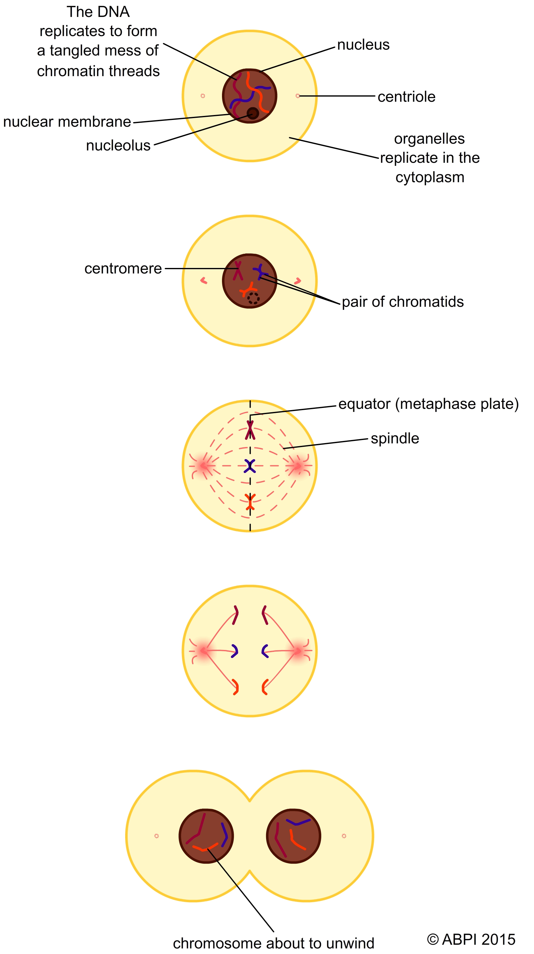
Nuclear division is an ordered process that results in the formation of two new cells with identical nuclei containing identical sets of chromosomes. Once mitosis starts it is a continuous process this can be seen by running the animation below without pauses. However to make it easier to understand, we usually describe it in stages.
Prophase: The pairs of chromosomes have replicated and become visible. Each chromosome is a pair of chromatids joined at the centromere. The nucleolus breaks down. The centrioles begin to separate, forming a spindle of microtubules between them.
Metaphase: The nuclear membrane begins to break down. The centrioles move to opposite poles of the cell and a spindle of microtubules stretches between them. The chromatids jostle and move to line up along the centre of the spindle (the equator or metaphase plate), each chromatid is attached to the spindle fibre by the centromere.
Anaphase: The centromeres between the chromatids separate. The proteins of the spindle fibres contract and the chromatids are pulled along the spindle fibres towards the opposite poles of the cell, with the centromeres leading.
Telophase: In early telophase the chromatids reach the poles of the cell and are now known as chromosomes again. New nuclear membranes start to form around each group of chromosomes. Nucleoli form in the new nuclei, and centrioles reform in both cells. The division of the cytoplasm begins, leading to cytokinesis.
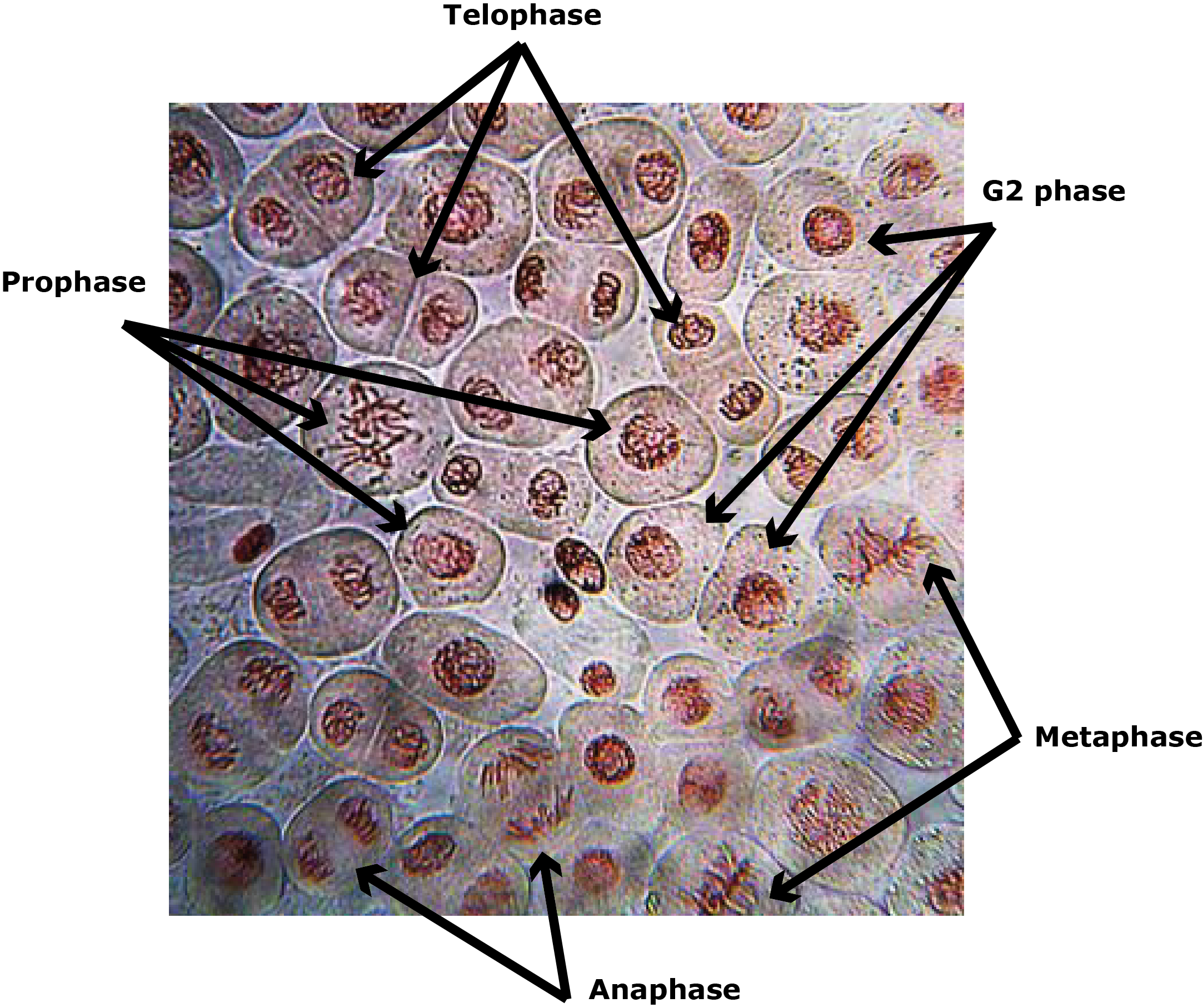
Microscopic images of onion root tip squash showing stages of mitosis.
(Photo credit: staticd. Licensed under the Creative Commons Attribution-Share Alike 3.0 Unported license)
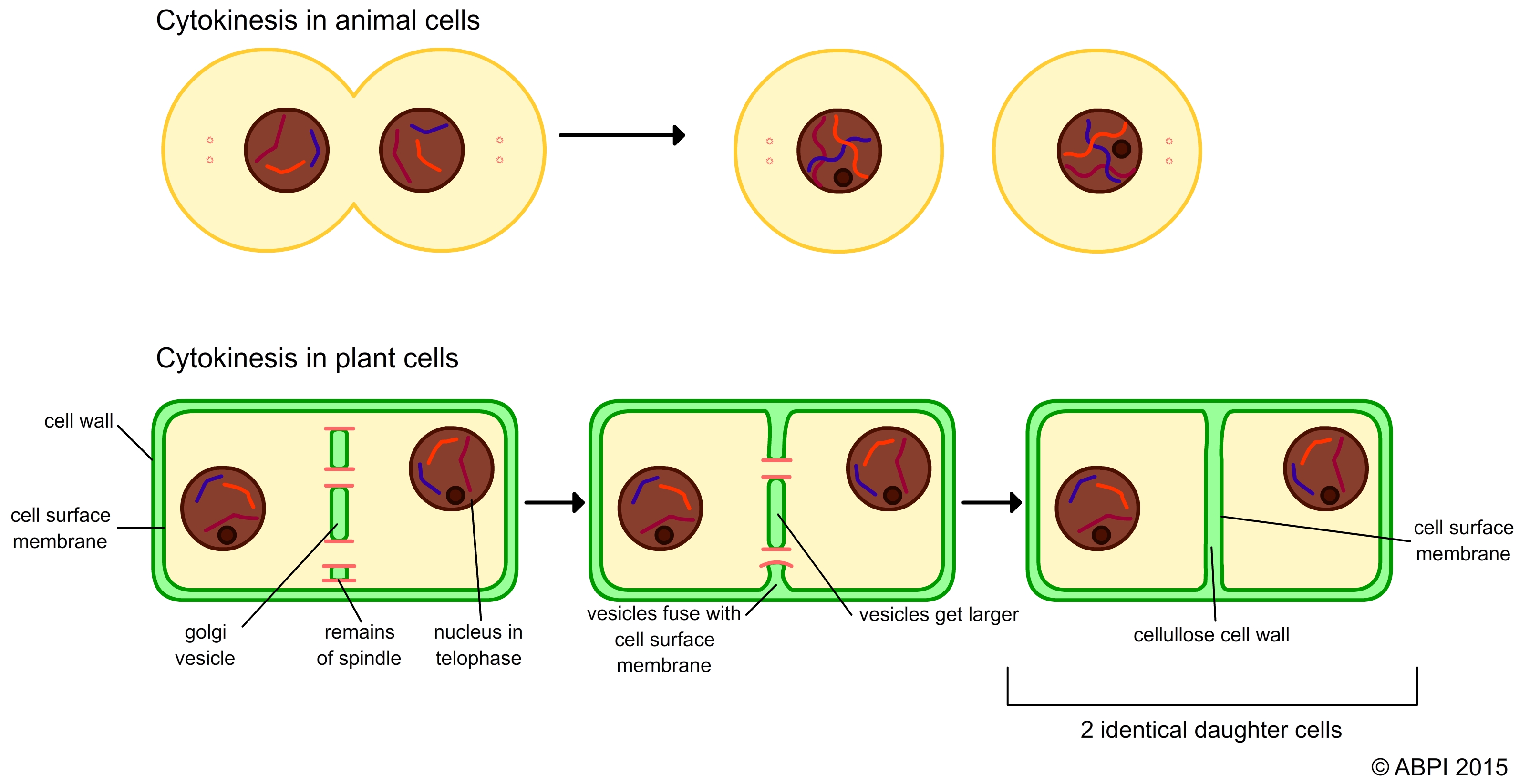
The final stage of cell division is the splitting of the cytoplasm along with all the organelles it contains. The cell surface membrane has to reform around each of the new cells. In plant cells, new cellulose cell walls also have to form and so the process is rather more complicated.
In animal cells
a ring of contractile protein fibres tighten around the middle of the cell like a belt, until the two halves are separated. The cell membrane is flexible and seals around the two halves.
In plant cells
the cell wall builds up from the centre of the cell. Some of the spindle fibre remains left in the cytoplasm seem to guide Golgi vesicles to line up in the middle of the large single cell with two nuclei. They form vacuoles that extend and fuse with the surface cell membranes, separating the contents of the two new cells. Cellulose plates are then laid down and fuse to form a complete cell wall, completing the two new cells.