This topic takes on average 55 minutes to read.
There are a number of interactive features in this resource:
 Biology
All of the millions of electrical impulses travelling around your body in the sensory and motor neurones are no use without some overall coordination. This is the role of the central nervous system, made up of the brain and the spinal cord.
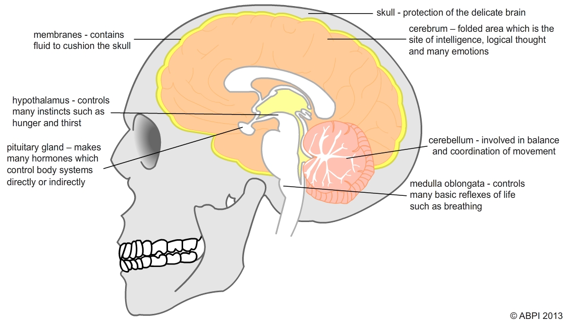
The brain is an organ made almost entirely from neurones, and it has the consistency of thick yoghurt. It is surrounded by membranes and protected from physical damage by the bones of the skull.
The human brain is a very complex structure. This single organ controls everything from our basic breathing and heart rhythm to our most complicated emotions and thought processes. Most of the brain is made up of grey matter – the cell bodies of neurones and the thousands of synapses which link them together. There is also white matter – the axons which lead into and out of the brain. Different areas of the brain are linked to different functions, shown in the diagram below:

The brain is not very easy to investigate. It is hidden away inside the skull. It is often only when the brain is damaged that we can appreciate exactly what it does and what an amazing organ it is. So if the right hand side of your brain is damaged, you may not be able to recognise even your closest family, while if the left side is damaged you may lose the ability to speak, even though you may still understand what other people say to you.
In the past, most of what we knew about the human brain was built up by studying what happened to people when their brain was damaged by accidents or by disease.
Phineas Gage was a foreman on a railway construction project in 1848. He was a well liked, calm and good mannered man who was respected by his colleagues. In a horrific accident, an explosion sent a metre long metal rod right through his skull, destroying part of the frontal lobes of his brain. Amazingly he survived and went on to live for another 12 years. However his personality seemed to have been affected.
Reports say that he became much more aggressive, used bad language and was much less inhibited in his behaviour after the accident. This gave doctors and scientists their first clear insight into the roles of specific areas of the brain – and the way it can compensate when part of the structure is lost. Gage’s skull and the tamping iron that went through it are on display at Harvard Medical School.

Today we no longer depend on the results of terrible accidents and disease to find out more about how the brain works. Scientists are beginning to find out exactly what happens in our brains as we react to the world around us, without needing to open up our skulls.
MRI scanners give doctors and scientists a way of seeing inside your body without having to open you up – and without needing to use X-rays which can be harmful. An MRI scanner uses radio waves and strong magnetic fields to produce images of the tissues and organs of your body – including the brain. Functional MRI (fMRI) scans are a relatively new way of taking detailed pictures inside the brain while you are doing something or feeling a particular emotion. The images show the areas where the blood flow increases or the amount of oxygen used by the tissues goes up. This tells scientists which areas of the brain are active as you look at or do different things.