Skip to content
    • Biology Biology
    • 16+
    • 14-16
    • 55

Cell biology

  of  10

Cell membranes

Cell membranes are vital to the way cells function. In animal cells, they form the outer layer of the cell – the ultimate barrier between the inside of the cell and its surroundings. In plant cells, the cell surface membrane is inside a relatively rigid cellulose cell wall but the properties of the membrane still control much of what moves into and out of the cell. Most of the organelles inside a eukaryotic cell are also membrane-bound. Understanding the properties of cell membranes is key to understanding how cells work.


The structure of the cell membrane

Our current model of the cell membrane has been built up over many years by a combination of experimental data and electron microscopy.


The unit membrane

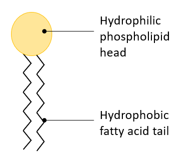
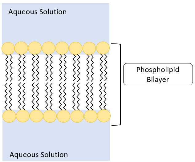
The basic structure of the cell membrane is a bilayer of phospholipids. Phospholipid molecules have a hydrophilic ‘head’ region around the ionic phosphate group and a long hydrophobic hydrocarbon tail. These polar lipids form a bilayer in aqueous solutions with the hydrophilic heads pointing outwards and the hydrophobic tails forming a hydrophobic layer in the middle. This bilayer is known as a unit membrane.

Individ. Phospholipid
Unit Membrane

The cell membrane

The cell membrane, however, is more than a simple unit membrane. Our current model is of a fluid phospholipid bilayer with many other molecules associated with it, floating or embedded in the lipid sea. These other molecules include cholesterol, glycolipids, proteins and glycoproteins and they all have different functions in the membrane. This is the fluid mosaic model of membrane structure and it explains many of the properties of membranes that we can observe experimentally.


Fluid Mosaic Model

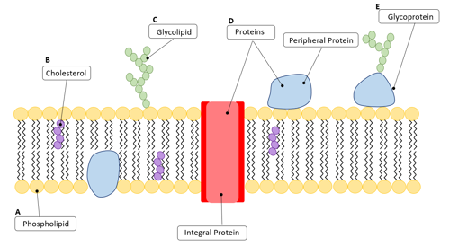
The fluid mosaic model of the cell membrane.


  • A Phospholipids: lipid molecules with a hydrophilic ‘head’ region around the ionic phosphate group and a long hydrophobic hydrocarbon tail that form a bilayer in aqueous solutions.
  • B Cholesterol: a lipid with a steroid ring structure, and hydrophilic and hydrophobic regions. It makes up part of the membrane structure - there may be up to one cholesterol molecule for every two phospholipids. Cholesterol makes the membrane stiffer and more rigid – so the amount of cholesterol in the structure affects the rigidity of the membrane.
  • C Glycolipids: lipids that have a carbohydrate chain attached to them. The carbohydrate chain is attached to the outside of the cell and is part of the cell recognition system.
  • D Proteins: a wide variety of molecules that carry out many of the very specific functions of the cell membrane. There are integral proteins and peripheral proteins. They can form temporary and permanent channels in the membrane, allowing different molecules to pass in and out of the cell. They may be enzymes involved in active transport systems or enzymes linked to biochemical pathways such as photosynthesis or respiration. Proteins also act as receptor molecules for other molecules such as hormones and neurotransmitters.
  • E Glycoproteins: proteins that have a carbohydrate chain attached to them. The carbohydrate chain sticks out of the outside of the cell and is part of the cell recognition system.


Functions of the cell membrane

Many of the functions of the surface cell membrane and membranes around cell organelles are similar, although there are some which are specific to the outer membrane.

  • Membranes form partially permeable barriers between the cell and its environment, between organelles and the cytoplasm and within organelles. They control the movement of substances both into and out of the cell and into and out of organelles. Permanent and temporary protein pores are involved in this control, as well as permanent and temporary active transport systems. Some channels are gated – they can be opened or closed depending on conditions inside or outside of the cell as described on the next page.
  • Membranes are the site of many chemical reactions because the enzymes involved are embedded in the membrane structure. Reactions take place both on the cell surface membrane and on the membranes in organelles such as mitochondria and chloroplasts.
  • Membranes are important in the development of chemical and electrochemical gradients – for example those involved in nerve impulses and in the production of ATP by chemiosmosis.
  • Membranes are the site of cell identification. The carbohydrate markers attached to glycoproteins and glycolipids along with some membrane proteins act as antigens, identifying one cell to other cells. For example this system enables the cells of the immune system to identify pathogens, cells from other organisms of the same species (eg after an organ transplant), abnormal body cells (eg cancer cells) and toxins produced by pathogens.
  • Membranes are the site of cell communication. Cell signalling takes place between cells through the protein receptor molecules in the cell surface membrane and within cells; for example in the passing of hormone messages from the body to the nucleus of the cell and the movement of mRNA out of the nucleus through nuclear membrane pores. This process is described in more detail later.

The pores in the nuclear membrane allow chemicals to move into the nucleus and mRNA to move out into the cytoplasm. (Image courtesy of Don W. Fawcett/Hector E. Chemes/Bernard Gilula (CC BY-NC-ND 3.0))

Activity

Use materials of your choice – anything from plasticine to plastic bottles and beyond – make a three-dimensional model of the cell membrane that can be used to explain the structure and functions of this amazing structure.